Download PDF
Natalie Allen
5th year medical student
University of Auckland
Abstract
A 15-month-old child presented to the emergency department with a popular acrally distributed erythematous rash of unknown cause. The eruption was unusually florid but consistent with a diagnosis of Gianotti Crosti syndrome, a self-limiting skin condition of childhood. The child had not been unwell, nor recently vaccinated, but tested positive for enterovirus polymerase chain reaction (PCR). Although numerous viruses, such as Epstein-Barr and Coxsackie B, have been associated with Gianotti Crosti syndrome, this is one of the few published cases specifically discussing the presentation of enterovirus-induced Gianotti Crosti syndrome.
Introduction
Gianotti Crosti syndrome (also called papular acrodermatitis) is a benign self-limiting skin condition of childhood. It usually presents as a monomorphous, purpuric rash, commonly appearing on the extensor surfaces of the extremities (1). Though frequently attributed to hepatitis B and Epstein-Barr virus, the syndrome is most often idiopathic (2). There are also European cases connecting atopy (3) as well as recent vaccination (4) with Gianotti Crosti syndrome. The pathogenesis of this condition is not well understood, but is likely immune-mediated and complex (3). This report hopes to draw attention to enterovirus as a cause of Gianotti Crosti syndrome, as well as a potential diagnosis for an unknown rash in children with no overt history of virus or recent vaccination.
Case Report
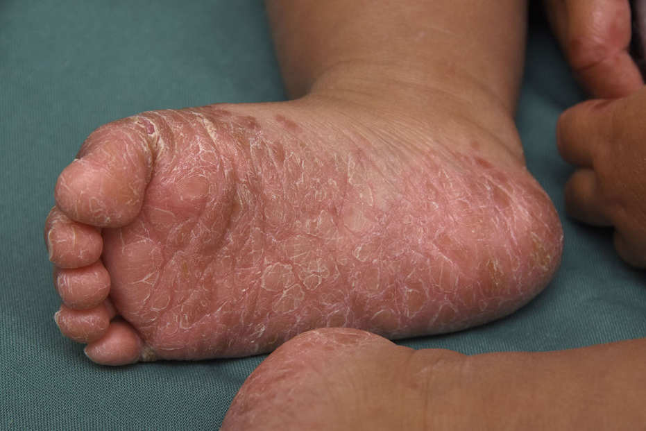
Figure 1. Bilateral foot rash
Rash as seen in dermatology clinic, two weeks after initial appearance. Desquamation and erythema still present despite hydrocortisone cream and amoxicillin use.
A 15-month-old male Māori infant presented to the Waikato Hospital Dermatology Department following referral from paediatrics. He had a two-week history of bilateral foot rash. The rash was initially managed in the community and emergency department with oral amoxicillin and hydrocortisone cream, which provided no benefit. Initially, the rash presented as erythematous desquamation of the feet, which then transformed into small white blisters. Following this, he developed papules on the extensor elbows, knees, buttocks, and around the mouth. He had no lymphadenopathy or mucosal involvement. There were no target lesions or intact vesicles. The child had a fever, but otherwise no coryzal or gastrointestinal symptoms. He had no history of recent illness or vaccinations in the last three months. He did have a history of upper respiratory tract infections, with two presentations to the emergency department at the age of 6 weeks and 5 months, alongside suspected dairy intolerance.
Differential diagnosis included acropustulosis of infancy, pityriasis rubra pilaris, and hand, foot, and mouth disease. His rash was most consistent with Gianotti Crosti syndrome. A complete blood count, antistreptolysin O, liver function enzymes, hepatitis and Epstein-Barr serology, and a throat swab were ordered. All results came back unremarkable except for the throat swab, which revealed presence of enterovirus. Management of Gianotti Crosti syndrome is conservative; the patient was prescribed fatty cream three times daily and followed up after one week. At this appointment, he improved significantly and was discharged from dermatology.
Discussion
Gianotti Crosti Syndrome was initially described in the 1950s in Rome and was previously thought to be associated with hepatitis B infection in children (6). A papular urticarial rash in a child lasting longer than ten days is considered the most predictive diagnostic indicator, as rashes consistent with Gianotti Crosti syndrome last between two and four weeks (5). The clinical features can be very variable, but usually include a symmetric papular eruption over the buttocks, feet, cheeks, and extensor surfaces of limbs. This rash may progress to vesicles and is often pruritic, though not in all cases. The incidence of Gianotti Crosti syndrome is difficult to predict, primarily due to underdiagnosis and the limited number of case studies. It is believed to occur predominantly in children between the ages of one and six years (6).
Despite the variety of potential causes, the pathogenesis of Gianotti Crosti syndrome remains unclear. The virus appears to be the key driver behind the skin changes, with secondary immunomodulation necessary to induce the rash (7). Despite using immunohistochemistry and electron microscopy, no viral antigens have been identified in the skin lesions of children presenting with Gianotti Crosti syndrome in at least three studies so far (7). The implications of this are that the development of the rash is unlikely due to local interactions between viral antigens and the skin, but rather some unknown systemic process (6).
The introduction of hepatitis B vaccination in 1965 challenged the etiology of this condition, and over time numerous additional causes were identified. Epstein-Barr virus is now considered to be the predominant precursor to Gianotti Crosti syndrome in the developed world (1). Other known viral agents include hepatitis A, cytomegalovirus, rotavirus, parainfluenza, mumps, coxsackie, and respiratory syncytial viruses (6). Bacterial causative agents such as Mycoplasma pneumoniae and haemolytic streptococci have also been reported (7). Vaccination has also been recorded as an inducer of Gianotti Crosti syndrome, though it has been speculated that afflicted children also had a viral infection at the time of immunisation (4,6). Alongside these infective cases, many cases are idiopathic (8). It also appears as though children with a history of autoimmunity are at a greater risk than their non-atopic peers (3).
Enterovirus has previously been included in the general list of causes (8). Regarding this specific case, the patient has no indications of enterovirus infection. This raises the question: how many idiopathic cases of Gianotti Crosti syndrome have an occult viral cause? The frequency of idiopathic presentation (8), coupled with asymptomatic viral infection discussed in this case may suggest that the development of Gianotti Crosti syndrome is not directly correlated with the severity of infection. A stronger understanding of the pathogenesis of this syndrome is necessary to understand how a variety of pathogens may cause similar presentations in young children. Therefore, this case report highlights the necessity for additional research with respect to the prevalence and presentation of enterovirus-induced Gianotti Crosti syndrome.
References
About the author
Natalie is a fifth year medical student at the University of Auckland. Her hobbies include overconsumption of cappuccinos and re-reading Harry Potter.
Acknowledgements and consent
Thank you to the Waikato Dermatology Department. Verbal and written consent were obtained from the patient's legal guardian.
Correspondence
Natalie Allen: [email protected]
A 15-month-old male Māori infant presented to the Waikato Hospital Dermatology Department following referral from paediatrics. He had a two-week history of bilateral foot rash. The rash was initially managed in the community and emergency department with oral amoxicillin and hydrocortisone cream, which provided no benefit. Initially, the rash presented as erythematous desquamation of the feet, which then transformed into small white blisters. Following this, he developed papules on the extensor elbows, knees, buttocks, and around the mouth. He had no lymphadenopathy or mucosal involvement. There were no target lesions or intact vesicles. The child had a fever, but otherwise no coryzal or gastrointestinal symptoms. He had no history of recent illness or vaccinations in the last three months. He did have a history of upper respiratory tract infections, with two presentations to the emergency department at the age of 6 weeks and 5 months, alongside suspected dairy intolerance.
Differential diagnosis included acropustulosis of infancy, pityriasis rubra pilaris, and hand, foot, and mouth disease. His rash was most consistent with Gianotti Crosti syndrome. A complete blood count, antistreptolysin O, liver function enzymes, hepatitis and Epstein-Barr serology, and a throat swab were ordered. All results came back unremarkable except for the throat swab, which revealed presence of enterovirus. Management of Gianotti Crosti syndrome is conservative; the patient was prescribed fatty cream three times daily and followed up after one week. At this appointment, he improved significantly and was discharged from dermatology.
Discussion
Gianotti Crosti Syndrome was initially described in the 1950s in Rome and was previously thought to be associated with hepatitis B infection in children (6). A papular urticarial rash in a child lasting longer than ten days is considered the most predictive diagnostic indicator, as rashes consistent with Gianotti Crosti syndrome last between two and four weeks (5). The clinical features can be very variable, but usually include a symmetric papular eruption over the buttocks, feet, cheeks, and extensor surfaces of limbs. This rash may progress to vesicles and is often pruritic, though not in all cases. The incidence of Gianotti Crosti syndrome is difficult to predict, primarily due to underdiagnosis and the limited number of case studies. It is believed to occur predominantly in children between the ages of one and six years (6).
Despite the variety of potential causes, the pathogenesis of Gianotti Crosti syndrome remains unclear. The virus appears to be the key driver behind the skin changes, with secondary immunomodulation necessary to induce the rash (7). Despite using immunohistochemistry and electron microscopy, no viral antigens have been identified in the skin lesions of children presenting with Gianotti Crosti syndrome in at least three studies so far (7). The implications of this are that the development of the rash is unlikely due to local interactions between viral antigens and the skin, but rather some unknown systemic process (6).
The introduction of hepatitis B vaccination in 1965 challenged the etiology of this condition, and over time numerous additional causes were identified. Epstein-Barr virus is now considered to be the predominant precursor to Gianotti Crosti syndrome in the developed world (1). Other known viral agents include hepatitis A, cytomegalovirus, rotavirus, parainfluenza, mumps, coxsackie, and respiratory syncytial viruses (6). Bacterial causative agents such as Mycoplasma pneumoniae and haemolytic streptococci have also been reported (7). Vaccination has also been recorded as an inducer of Gianotti Crosti syndrome, though it has been speculated that afflicted children also had a viral infection at the time of immunisation (4,6). Alongside these infective cases, many cases are idiopathic (8). It also appears as though children with a history of autoimmunity are at a greater risk than their non-atopic peers (3).
Enterovirus has previously been included in the general list of causes (8). Regarding this specific case, the patient has no indications of enterovirus infection. This raises the question: how many idiopathic cases of Gianotti Crosti syndrome have an occult viral cause? The frequency of idiopathic presentation (8), coupled with asymptomatic viral infection discussed in this case may suggest that the development of Gianotti Crosti syndrome is not directly correlated with the severity of infection. A stronger understanding of the pathogenesis of this syndrome is necessary to understand how a variety of pathogens may cause similar presentations in young children. Therefore, this case report highlights the necessity for additional research with respect to the prevalence and presentation of enterovirus-induced Gianotti Crosti syndrome.
References
- Marcassi AP, Piazza CA, Seize MB, Cestari SD. Atypical Gianotti-Crosti syndrome. An Bras Dermatol. 2018 Mar;93(2):265-7.
- Taieb A, Plantin P, Pasquier PD, Guillet G, Maleville J. Gianotti‐Crosti syndrome: a study of 26 cases. Edinb Med J. 1986 Jul;115(1):49-59.
- Ricci G, Patrizi A, Neri I, Specchia F, Tosti G, Masi M. Gianotti-Crosti syndrome and allergic background. Acta Derm Venereol. 2003 May 1;83(3).
- Andiran N, Şentürk GB, Bükülmez G. Combined vaccination by measles and hepatitis B vaccines: a new cause of Gianotti-Crosti syndrome. Dermatology. 2002;204(1):75-6.
- Chuh AA. Diagnostic criteria for Gianotti-Crosti syndrome: a prospective case-control study for validity assessment. Cutis. 2001 Sep;68(3):207-13.
- Brandt O, Abeck D, Gianotti R, Burgdorf W. Gianotti-crosti syndrome. Dermatology. 2006; 55(1):136-45.
- Baldari U, Monti A, Righini MG. An epidemic of infantile papular acrodermatitis (Gianotti-Crosti syndrome) due to Epstein-Barr virus. Dermatology. 1994;188(3):203-4.
- Snowden J, Badri T. StatPearls [Internet]. StatPearls Publishing, 2017. Acrodermatitis, papular (Gianotti Crosti syndrome). [cited XXX]. Available from: https://www.ncbi.nlm.nih.gov/books/NBK441825
About the author
Natalie is a fifth year medical student at the University of Auckland. Her hobbies include overconsumption of cappuccinos and re-reading Harry Potter.
Acknowledgements and consent
Thank you to the Waikato Dermatology Department. Verbal and written consent were obtained from the patient's legal guardian.
Correspondence
Natalie Allen: [email protected]


